Lower Back Muscle And Tendon Diagram / Lower Middle Back A Step Beyond Massage Therapy - Symptoms of muscle pain include:. These structures work together to support the body, enable a range of movements, and send messages from the brain to. The calf muscle, on the back of the lower leg, is actually made up of two muscles: As you can see, there are also have a spine of scapula deltoid, triceps brachii, latissimus dorsi. They help support particular bones and make them move. Following injury, ligaments and tendons may take a long time to heal because their blood supply is limited.
Muscles of the lower back and buttocks diagram, human muscles, muscles of the lower back and buttocks diagram. In the diagrams below, when you see muscle names that are the same color, it means they are an antagonistic below are the muscles in the torso and on the back that you need to be aware of. But, they are common in the back and can cause pain. The pubis, ischium, and ilium together constitute the pelvis while the thigh bone is the femur. .lower back muscle diagrams labeled, muscle diagram of lower back, human muscles, human related posts of lower back muscle diagram.
Low Back Pain Pictures Symptoms Causes Treatments from img.webmd.com Muscles of lower leg (calf, soleus). Lower back muscle diagram anatomy does degenerative disc disease affect the lower back muscle? These muscles are located on the front side of the lower leg. Ligaments and tendons are fibrous tissues made up of collagen fibers. But, they are common in the back and can cause pain. Ligaments provide stability of the spine during movement and rest. No matter which one you have, the treatment is the same for. As you can see from the diagram to the right, there are many muscles and tendons that make up the hip and buttocks region.
Ligaments and tendons are fibrous connective tissues made up of densely packed collagen fibers.
The back consists of the spine, spinal cord, muscles, ligaments, and nerves. Muscle or ligament strains can occur from repeated use of the muscles, or from improperly or awkwardly lifting heavy objects. The muscles of the lower back help stabilize, rotate, flex, and extend the spinal column, which is a bony tower of 24 vertebrae that gives the body structure and houses the spinal cord. The muscles of the abdomen, lower back, and pelvis are separated from those of the chest by the muscular wall of the diaphragm, the critical breathing muscle. Ligaments and tendons are fibrous tissues made up of collagen fibers. The diagram shows the posterior (rear) view of the buttock. The back is a complex structure of bone and muscle, supported by cartilage, tendons and ligaments, and fed by a network of blood vessels and nerves. Another common cause of lower back and hip pain is disc injury. They help support particular bones and make them move. This is a diagram of the larger and more surface muscles of the low back. The 3 major spinal ligaments are the (1) anterior longitudinal ligament, (2) posterior longitudinal ligament and (3) ligamentum flavum. The muscles, bones, ligaments, and tendons in the back can all be injured and cause back pain. The gastrocnemius is the larger calf muscle, forming the bulge visible beneath the skin.
12 photos of the muscles of the lower back and buttocks diagram. The diagram shows the posterior (rear) view of the buttock. These muscles are located on the front side of the lower leg. Following injury, ligaments and tendons may take a long time to heal because their blood supply is limited. Low back supported by lumbar ligaments, tendons and muscles.
Lumbar Spine Anatomy from cloud2.spineuniverse.com They help support particular bones and make them move. As you can see from the diagram to the right, there are many muscles and tendons that make up the hip and buttocks region. Muscle spasms (contraction or stiffening of the back muscles) muscles that feel tight; This picture also contains humerus, olecranon process of ulna, deep to tendon and so on. Muscle or ligament strains can occur from repeated use of the muscles, or from improperly or awkwardly lifting heavy objects. Your lower back (lumbar spine) is the anatomic region between your lowest rib and the upper part of the buttock. As you can see, there are also have a spine of scapula deltoid, triceps brachii, latissimus dorsi. The 3 major spinal ligaments are the (1) anterior longitudinal ligament, (2) posterior longitudinal ligament and (3) ligamentum flavum.
As you can see from the diagram to the right, there are many muscles and tendons that make up the hip and buttocks region.
A strain can be an injury to a tendon attachment from muscle to bone. When the tissues are stretched too far, or torn, microscopic tears of varying degrees can occur. Systems of strong fibrous bands of ligaments hold the vertebrae and discs together and stabilize the spine by helping to prevent excessive movements. The most common type of back pain is muscle pain—also called muscle strain or soft tissue strain. To learn more about the anatomy of the spine, watch this video. The muscles of the abdomen, lower back, and pelvis are separated from those of the chest by the muscular wall of the diaphragm, the critical breathing muscle. Your lower back (lumbar spine) is the anatomic region between your lowest rib and the upper part of the buttock. Lying exposed between the protective bones of the superiorly located ribs and the inferiorly located pelvic girdle, the muscles of this region play a critical role in protecting the. #muscles of the lower back and hip diagram. Ligaments provide stability of the spine during movement and rest. Muscles of the lower limb. The gastrocnemius is the larger calf muscle, forming the bulge visible beneath the skin. Back pain rarely gets the tendonitis diagnosis, but it's important to understand the tendonitis dynamic because that is the cause of back pain.
The back—especially the lumbar, or lower back—bears much of the body's weight during walking, running, lifting and other activities. Lower back muscle and tendon diagram : The most common type of back pain is muscle pain—also called muscle strain or soft tissue strain. Human muscle system, the muscles of the human body that work the skeletal system, that are under voluntary control, and that are concerned with movement, posture, and balance. Muscles of the hip & thigh (quadriceps, hips).
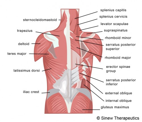
Lower Back Pain Strain Information Sinew Therapeutics from sinewtherapeutics.com Back pain rarely gets the tendonitis diagnosis, but it's important to understand the tendonitis dynamic because that is the cause of back pain. As you can see, there are also have a spine of scapula deltoid, triceps brachii, latissimus dorsi. The back—especially the lumbar, or lower back—bears much of the body's weight during walking, running, lifting and other activities. When the tissues are stretched too far, or torn, microscopic tears of varying degrees can occur. Ligaments and tendons are fibrous connective tissues made up of densely packed collagen fibers. #muscles of the lower back and hip diagram. Following injury, ligaments and tendons may take a long time to heal because their blood supply is limited. Lying exposed between the protective bones of the superiorly located ribs and the inferiorly located pelvic girdle, the muscles of this region play a critical role in protecting the.
In the back and elsewhere in the body, tendons attach muscles to bones.
Muscles of the lower limb. • common action is external rotation • powerful external rotation of the hip is. Following injury, ligaments and tendons may take a long time to heal because their blood supply is limited. This is a diagram of the larger and more surface muscles of the low back. The 3 major spinal ligaments are the (1) anterior longitudinal ligament, (2) posterior longitudinal ligament and (3) ligamentum flavum. It connects the sacrum (lower region of the spine) to the top of the femur (thigh bone) and aids in external rotation (turning out) of the hip joint. Evenly distribute weights from your upper body into the lower extremities. To learn more about the anatomy of the spine, watch this video. Muscles of lower leg (calf, soleus). 12 photos of the muscles of the lower back and hip diagram. Back pain is one of the most common kinds of pain. A strain can be an injury to a tendon attachment from muscle to bone. This curve, called lordosis, helps to:
There are around 650 skeletal muscles within the typical human body lower back muscle diag. Lower back muscle and tendon diagram :

0 Comments:
Posting Komentar